Although the diagnosis and treatment options of oncology patients are improving worldwide, malignant tumour-related mortality remains high. This is due to the resistance of the tumour, – its ability to survive after various therapies. As explained by Zane Simsone, a cell biologist, researcher at the University of Latvia and manager of the project “Development of resistance as a result of tumour therapy” administered by the UL Foundation, the therapy is not always successful due to tumour heterogeneity, cancer cell polymorphism and different cell functional activity within the same cancer tissue. Namely, cancer cells are heterogeneous, furthermore, they have genetically different forms.
The latest data and results obtained by the UL ICRM research group will help to describe microcells in detail and to establish when they occur, when they are able to absorb the greatest volume of substances, moreover, researchers will explore other features of microcells. Such a study in future will contribute to clarify the reasons why tumour therapy is not always successful. In addition, the knowledge gained could be used to evaluate the effectiveness of anticancer therapies, and in a longer perspective serve as a basis for development of anticancer drugs. The project is supported by the platinum patron of the University of Latvia “Mikrotīkls” in cooperation with the University of Latvia Foundation.
Microcells – a threat to successful tumour therapy
Studies implemented worldwide suggest that cancer stem cells are resistant to the applied anticancer treatments. Chemotherapy and radiation therapy, which are extensively used in practice, cause DNA damage, preventing tumour cells from dividing and promoting their death. Although cancer stem cells are associated with low efficacy of therapy, they are referred to as a heterogeneous tumour subpopulation. Studies carried out to date have shown that cancer stem cells have mechanisms for restoring DNA, thus being able to avoid the impact and effects of medication. There are several hypotheses about cancer stem cells, and they show the following main features of cancer stem cells:
- only a small number of tumour cells have tumorigenic (tumour-forming) potential;
- the population of cancer stem cells can be distinguished from other cancer cells by their surface markers;
- the cancer stem cell population has the potential for self-renewal and proliferation.
Cancer stem cells are not considered after the applied therapy, respectively, cancer-resistant cells and their properties are not described. In Latvia, research on cell changes and formation by looking at tumour cell lines under the influence of anticancer therapy is being studied by several scientists. Currently, the only effort to study microcells is done by Doctor of Biology, scientist Indulis Buiķis and his colleagues. The research team studies small cells – microcells. These are round or oval cells with a small cytoplasm (the parts of the cell that cover the nucleus). Microcells are described as natural components of the tumour, and their number increases under the influence of applied anticancer therapies, i.e., microcells are considered to be potential tumour regenerating cells after the death of the tumour itself. The first results show that intensive biochemical processes take place within the microcells. Such microcells have been observed, for example, in HT-1080 cell lines of Djungarian hamster and human sarcoma or malignant connective tissue tumour after treatment of the cells with anticancer drugs, methanol, hyperthermia treatment of the cells and growth of the cells in a cell culture medium containing inadequate nutrients.
Microcell formation is explained by the process of sporosis, when under the influence of anticancer therapy, new cells with stem cell-like properties may develop from fatally damaged cancer cells, resulting in the formation of one or more microcells from the damaged cancer cell.
1.  2.
2. 
3.  4.
4. 
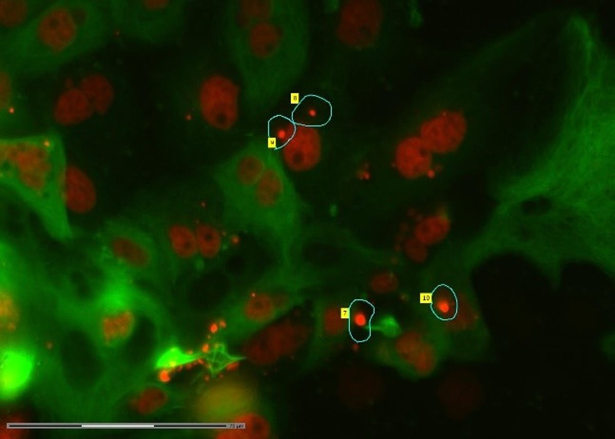
[1] Control cells of the porcine kidney cell line LLCPK1. Green – cytoplasm, red – nucleus. [2] Cells of the porcine kidney cell line LLCPK1 treated with an anticancer agent. Green – cytoplasm, red – nucleus. [3] Cells of the porcine kidney cell line LLCPK1 treated with an anticancer agent. Cell group marking for microdissection. [4] Cells of the porcine kidney cell line LLCPK1 treated with an anticancer agent. Marking of a single cell for microdissection. Image acquisition and cell isolation were performed with Laser Microdissection with ZEISS PALM MicroBeam. Author – Zane Simsone.
Visual control is a positive step in improving tumour therapy
The currently identified characteristics of microcells are their small size and endocytosis (ability to absorb other surrounding substances in the cell). The main difference between microcells and cancer stem cells is their ability to do endocytosis. Therefore, it is important to research therapy-resistant cells, determining their functional properties and markers characterising microcells. At present, the identification of microcells after the applied therapy is performed using morphological microscopy, immunohistochemical identification methods and flow cytometry, classifying the cells according to their size and expression markers. There are both advantages and disadvantages to using flow cytometry. The advantages – a larger number of cells can be analysed. However, a negative aspect is that the microcells in early development are smaller than 1.5 micrometres. Using standard flow cytometry, such samples fall below the detection threshold as artifacts. Thus, visual control would be important for single cell analysis, and this is provided by a microdissection device. Using this equipment, it is possible to obtain cells of interest to a research group for further analysis from cell cultures, sections of frozen tissue samples, and formalin-fixed paraffin-embedded tissue sections.
If the cells of interest are isolated from the cell culture, then further culturing of the isolated cells is possible to assess their viability. This summer, in collaboration with the ZEISS Microscopy Customer Centre in Oberkochen, Germany, project manager Zane Simsone, Dr. biol. Tālivaldis Freivalds and other colleagues of UL ICRM performed a pilot experiment with microcellular isolation after treatment with an anticancer agent and their regeneration. During the project, two cell lines were used – human osteosarcoma (malignant bone tumour) and porcine kidney cell lines. Cells were treated with an anticancer agent, and after two days, subpopulations of various cells, including microcells, were isolated using a microdissection device. The isolated cells were further cultured until day 7, with a photograph taken every second day. The research team continues to analyse the obtained data, and the current results already indicate that this method can be used to isolate the cell or group of cells of interest without damaging them.
About the University of Latvia Foundation
Since 2004, the UL Foundation provides an opportunity to patrons and cooperation partners to support both the University of Latvia and other leading higher education institutions of our country, thereby investing in the future of Latvia. The priorities of the UL Foundation are to support the most outstanding, dedicated students and researchers, to advance creation of a modern study environment, as well as to promote construction and refurbishment of university’s buildings.

 Academic Centre